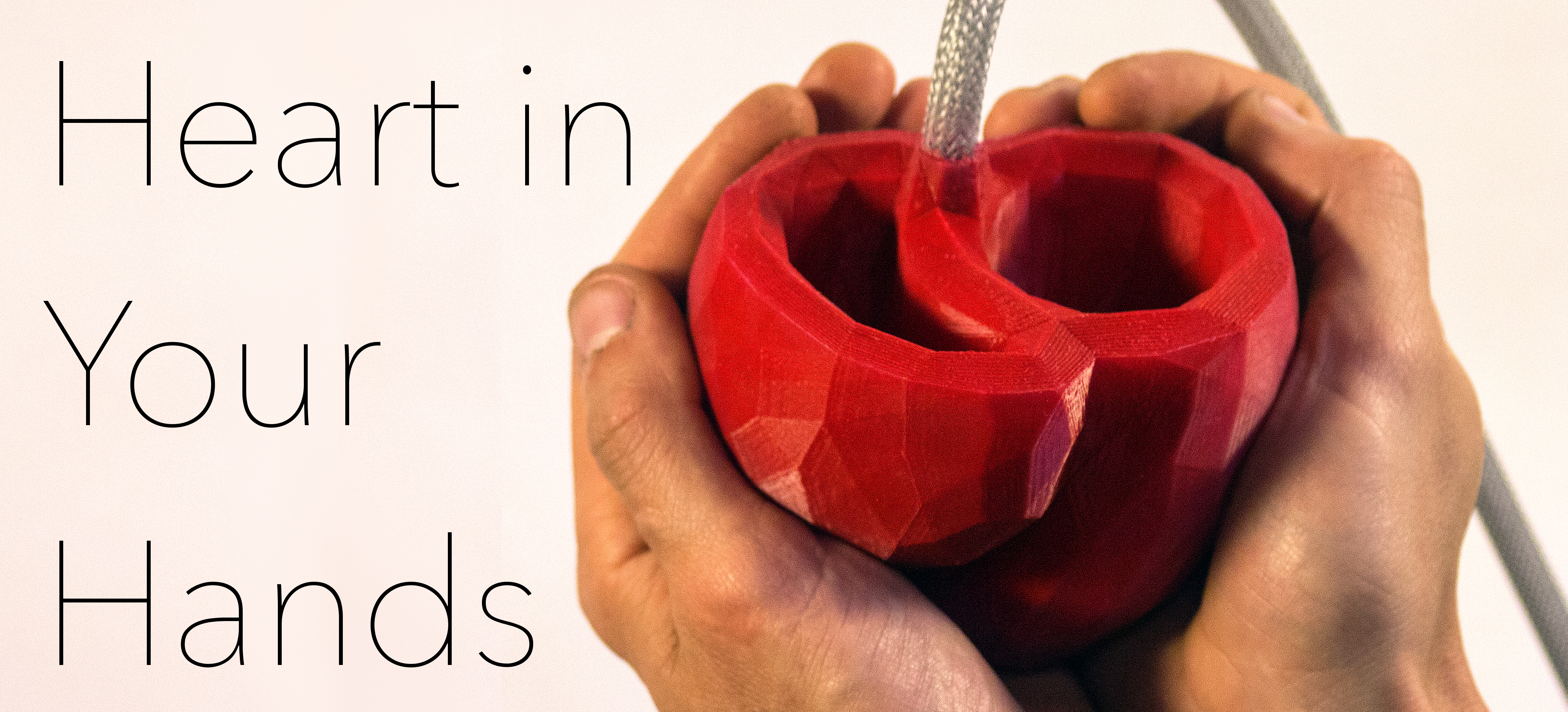
What is the Heart in Your Hands Exhibition?
Heart in your Hands is a collaborative effort between researchers at King’s College London, Rusty Squid, the Royal Academy of Engineers and the British Heart Foundation aimed at sharing the with you how the beating heart works, how disease can influence your heart, and how computational modelling and medical imaging are providing new avenues for personalised and predictive medicine.
What will I see at the Exhibition?
Heart in Your Hands takes you on a journey of discovery, showing you how the heart works and how research is improving our understanding of heart function in disease. This exhibition is built around three interactive displays to give you a glimpse into these exciting research areas and your own heart!

What is the Heart in Your Hands Exhibition?
Heart in your Hands is a collaborative effort between researchers at King’s College London, Rusty Squid, the Royal Academy of Engineers and the British Heart Foundation aimed at sharing the with you how the beating heart works, how disease can influence your heart, and how computational modeling and medical imaging are providing new avenues for personalized and predictive medicine.
What will I see at the Exhibition?
Heart in Your Hands takes you on a journey of discovery, showing you how the heart works and how research is improving our understanding of heart function in disease. This exhibition is built around three interactive displays to give you a glimpse into these exciting research areas and your own heart!

HEARTVIEWER
In this interaction, you will get a glimpse at the living heart through magnetic resonance images. Pick up one of our 3D model hearts and place it on our scanner to view how that heart beats, how the muscle of the heart moves, and how the blood flows into and out of its chambers. Compare between different hearts to see how medical imaging provides a novel noninvasive path for understanding the impact of heart function on performance and disease. Click here for more.
CARDIOSYNC
Re-pace our virtual heart and learn how computational modelling is changing the way we think about treatment. Pick up our heart controller, move it around and explore how abnormal heart rhythm affects the beating of the human heart. Choose from a selection of different pacing sites, and see how cardiac resynchronisation therapy can restore electrical rhythm and how computational models provide a way to predict the effects of therapy. Click here for more.
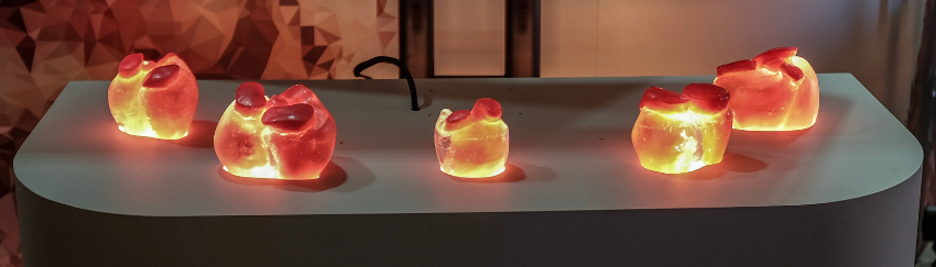
HEART IN YOUR HANDS
Ever wonder how the heart beats? How about how the heart beat changes with disease? Explore the basic biomechanics of the heart through our soft robotic heart models. Paired to a wearable heart rate monitor, you can watch our soft robotic hearts beating in time with your own. Pick up our dilated cardiomyopathy model, and experience first hand how heart failure changes the size, shape and beating of the heart. Click here for more.
How does the Human Heart Work?
Each beat of your heart relies on a hierarchy of structure and function. Explore the diagram below to learn how fundamental proteins and ions within our cells drive the contraction of the heart and push blood through the body.
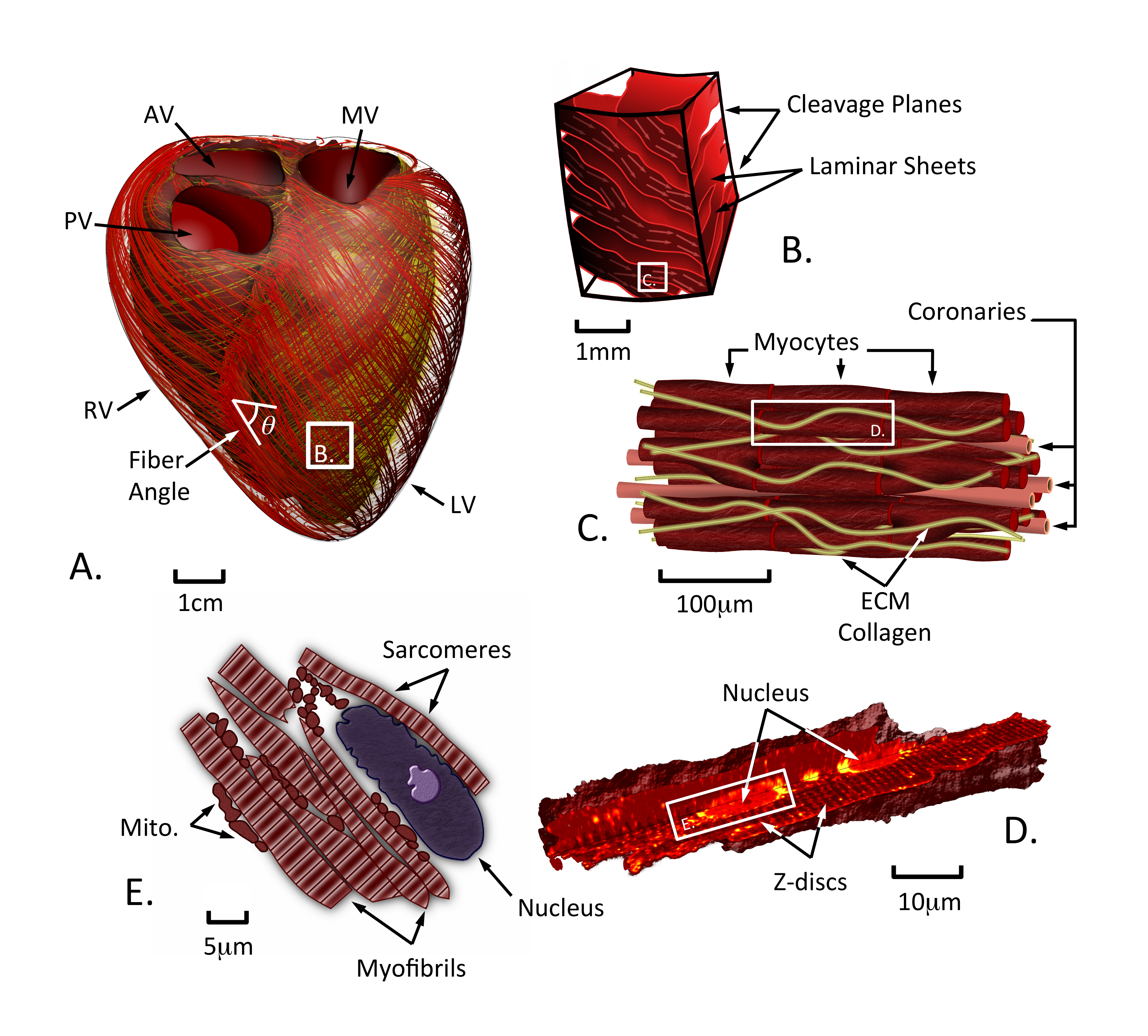
(A) Your heart consists of four chambers, divided into thin-walled atria and thick-walled ventricles (shown). Your heartbeat is caused by the propagation of an electrical wave that moves across the atria, propagates through the purkinje fibre network and through the muscle. Electrical stimulation causes the shortening of muscle fibre conglomerates (shown as red and yellow lines). This shortening compresses the ventricular chambers causing blood to be pushed through the cardiovascular system.
(B) To effectively squeeze its chambers, the heart must coordinate fibre shortening. This is done regionally in the heart by organising fibres together into laminar sheets that can contract, creating thickening of the heart wall. Sheets are separated by cleavage planes that allow the muscle to thicken more effectively.
(C) Within each laminar sheet, the heart muscle is comprised of fibres and blood vessels held together by a strong scaffolding known as the extracellular matrix. Fibres are composed of long muscle cells, known as myocytes, that are strung end-to-end to enable the propagation of the electrical wave through the tissue and fibre shortening. Capillaries run throughout the muscle to provide energy to the myocytes (metabolites) and clear out waste.
(D&E) Each myocyte is packed tight with myofibrils. Myofibrils are composed of sarcomeres – the fundamental contractile unit of the cell – separated by Z-discs. Electrical excitation of the myocyte causes an increase in the number of calcium ions free within the cell, unlocking proteins within the sarcomere that enable them to contract. Energy for contraction stems from breaking the molecule adenosine triphosphate (ATP), releasing energy enabling movement of sarcomere proteins. ATP is principally created within mitochondria found throughout the myocyte.
Research in the fight against Heart Failure
From new drugs and devices, new imaging techniques, and computer-based technologies, research is paving the way for innovation in healthcare. Funded by charities like the British Heart Foundation and national research councils like the EPSRC, engineering research efforts are continuing to be developed and refined in our efforts to fight heart failure. Our exhibition focuses on two core research technologies.
COMPUTATIONAL MODELLING
Computational modeling is a tool enabling the simulation of complex phenomena, like the beating of the human heart. Noting that certain quantities — such as momentum, energy, or charge — are conserved, mathematical equations can be derived that explain how a system will evolve. For example, conservation of momentum principles provide equations (partial differential equations) telling us how tissue will move or blood will flow when exposed to different pressures.
These mathematical equations are often too complex to solve with a pen and paper. Instead, they are approximated using numerical techniques that simplify the equations into a form that can be solved using your computer. This approach enables engineers to take medical images from a patient, construct a computer model of that patient’s heart, and use fundamental physics to learn about the function of that heart and how it may respond to treatment. For more on how computational modelling is changing our view of the heart, click here.
MAGNETIC RESONANCE IMAGING
Magnetic Resonance Imaging (MRI) is a technique used in clinics to acquire pictures of the anatomy and function within the body. Cardiac MRI focuses on creating images within the heart and can be used to observe alterations in heart anatomy, structure and motion. Because of its clear resolution of different tissues, MRI is a popular technique for examining many different cardiovascular conditions.
While MRI has proven to be effective for clinical assessment of your heart, research is continually pushing the envelope of what can be observed in the heart. Techniques like MR elastography provide a means for assessing the stiffness of muscle tissue. New advancements in acquisition and analysis are speeding up the pace of scans and providing more clear and consistent data. MRI research is making it possible to see regions of scar tissue, restructuring of the muscle, and poor activation of the heart itself. For more on how magnetic resonance imaging works, click here.
After the Exhibition?
Has the exhibition piqued your interest? Keep up-to-date with the research and developments within our department through our website and blog, as well as the amazing displays created by Rusty Squid. Get involved by considering some of our teaching and research programmes, including our Centre for Doctoral Training in Medical Imaging.
Interested in volunteering or donating to help in the fight against Heart Failure? Team up with the British Heart Foundation in their efforts to improve care for the over half a million with heart failure.